Impact Of Arterial Flow Complexity On Flow Diverter Outcomes

The flow diverter is becoming a standard device for treating cerebral aneurysms. The aim of this in vitro study was to evaluate the impact of flow complexity on the effectiveness of flow diverter stents in a cerebral aneurysm model. The flow pattern of a carotid artery was decomposed into harmonics to generate four flow patterns with different pulsatility indexes ranging from 0.72 to 1.44. The effect of flow diverters on the aneurysm was investigated by injecting red dye or erythrocytes as markers. The recorded images were postprocessed to evaluate the maximum filling of the aneurysm cavity and the washout time. There were significant differences in the cut-off flows between the markers, linked to the flow complexity. Increasing the pulsatility index altered the performance of the flow diverter. The red dye was more sensitive to changes in flow than the red blood cell markers. The flow cut-off depended on the diverter design and the diverter deployment step was crucial for reproducibility of the results. These results strongly suggest that flow complexity should be considered when selecting a flow diverter.

Effect of placement on mesh density of the flow diverter. Representative images of the aneurysm cavity at time 0 and 20 s during the 1st , 2nd , and 3rd placement of the flow diverter (porosity 44.2%) are shown. The middle panels are enlargements of the 1eft panels.
Flow patterns applied to the aneurysm. Four different target flow patterns were considered for the system (dashed lines). These included the first harmonic (a), the 1st to 3rd harmonics (b), the 1st to 5th harmonics (c), and the 1st to 15th harmonics (d) from the reference carotid flow, namely H1, H3, H5, and H15 respectively. The resultant effective experimental flow rate for each pattern is shown as a solid line. The pulsatility index of the measured flow is indicated in brackets.

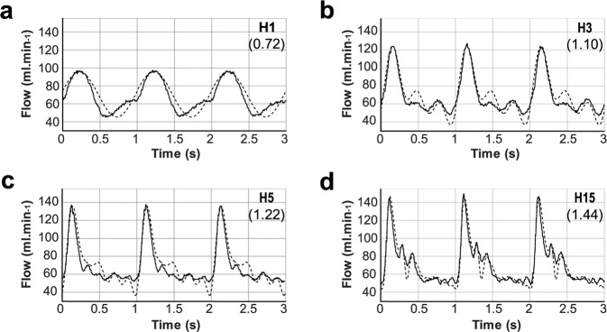
Four flow patterns were applied as input flow to a silicone model of a cerebral aneurysm of 10 mm diameter and 1.1 aspect ratio, which was therefore considered a large aneurysm. A carotid artery mean flow was used. The flow patterns H1, H3, and H5 were modelled by summing the 1st, 3rd, and 5th harmonics of the reference flow. Information on the flow complexity was gathered over the first 15 harmonics and the H15 flow pattern showed less than 1% variation from the original flow pattern.
Although the minimal flow rate was similar between these patterns (47 to 52 ml.min−1), the peak flow rate increased from the H1 to the H15 patterns (97 to 146 ml.min−1); consequently, the measured pulsatility indexes for H1, H3, H5, and H15 reached 0.72, 1.1, 1.22, and 1.44, respectively. Increasing the number of harmonics included in the input signal also increased the number of peaks during one cardiac cycle, sharpening these peaks. The observed maximal acceleration of the flow rate ranged from 350 ml.min−2 to 1500 ml.min−2 for H1 to H15, respectively.

Scroll to Top